Depending on appearance and histological structure, scars are divided into normotrophic, atrophic, hypertrophic and keloids. The last to types refer to the group of pathological scars.
Normotrophic scars
Normotrophic scars are leveling with the surrounding skin are the most harmless by nature (scar removal techniques). They are formed as a result of an adequate reaction of the organism to the injury. Later they become thin, whitish and do not cause physical discomfort to the bearer. Such scars typically do not require correction, apart from the cases of aesthetical improvement of scar appearance or acceleration of colour normalization process (scars treatment Stockholm)
Atrophic scars
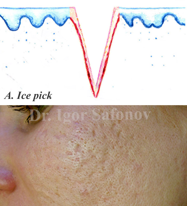
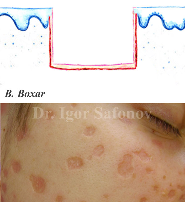
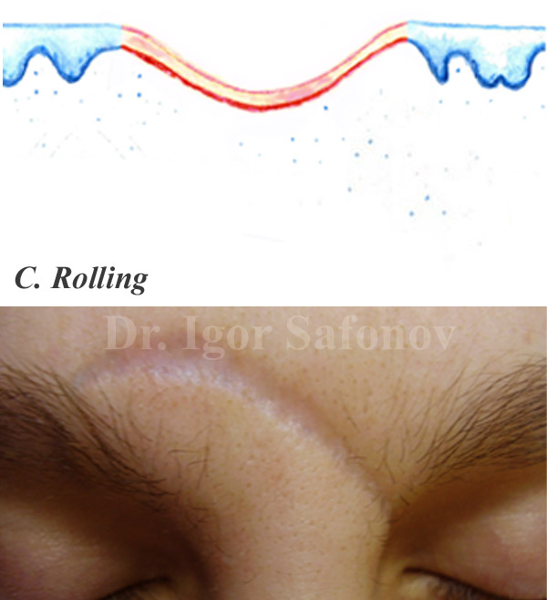
Atrophic scars are below the skin level are often the result of injury or inflammation (atrophic scar reduction treatment). Atrophic scars are subdivided into the types as follows: ice pick scars (Fig.2.A); boxcar scars (Fig.2.B) and rolling scars (Fig.2.C)
Fig.2. Types of Atrophic Scars
Ice-pick scars are wedge-shaped or prickly occur exclusively after acne inflammation. They look like small deep holes on the skin surface after puncture with a sharp object. Ice-pick scars are wider on the skin surface and taper to the deep skin level.
Boxcar scars – round or oval indentations or craters in the skin (pits or sunken scars). Boxcar scars have sharp vertical edges and are similar to the scars left after chickenpox. They can be shallow or deep punched out wide at the surface and at the base. They are most often placed on the cheeks and temples.
Rolling scars are rolled semi-circular scarring that can occur as a result of acne healing process, injury or surgery. They appear as indentations in the skin and tend to measure a few millimeters wide. They are defined according to their sloping edges, which give the skin a wavy, uneven appearance. Rolling scars in acne are not always the same size as their size depends on how the skin heals. Rolling scars are more common in facial areas where the skin is thicker, such as the lower cheeks and jaw.
The skin over atrophic scars is in most cases thin and weak. It has linear transverse striations. Atrophic scars look pale because they lack pigment. This characteristic appearance is due to defective connective tissue under the scar, collagen and elastin deficiencies, the main proteins that form the structure of the skin. See also atrophic scars and acne scar treatment in Stockholm.
Keloid and hypertrophic scars
Keloids and hypertrophic scars are united here in the same section because it is very difficult to distinguish these scars in the early phases of their formation. During further maturation process of scars, the differences between keloids and hypertrophic scars become more clear. Therefore, treatment approach should also be modified according to their types.
Hypertrophic scar
A hypertrophic scar has raised scar bases that are placed above the skin surface. The difference between a hypertrophic scar and keloid is the growth pattern. A hypertrophic scar grows in height (Fig.3), but a keloid grows in width. Hypertrophic scars after breast surgery and caesarean section occur most often. They can also form after injuries due to severe inflammation, also as a result of associated secondary infection when the immune system is impaired, endocrine dysfunctions and others (Fig.3). See more Keloids and hypertrophic scars treatment, removal and correction.
Keloids (keloid scars)
Keloids significantly exceed the size of the original wound. Keloids after piersing is the main reason to occur in the ears region. By a keloid is observed massive proliferation of connective tissue in the areas of burns, wounds, post-acne (acne keloid scars) and post-surgery injuries. Collagen fibres in keloid scars are located chaotically and node-like, resembling a fingerprint (Fig.4) Despite their age, keloids can be active (growing) and inactive (stabilized). An active keloid scar grows and causes pain, itching, numbness, emotional distress, and looks like a strained red scar, often with a bluish tint. Inactive keloid is not growing and does not bother the bearer from the subjective perspective, has a pink colouring or the one close to the colour of normal skin. Keloids are typically localized in the regions of ear lobe, decollate (chest), shoulders and back (Hypertrophic scars and keloids before and after treatment).


